- 移动端


爱必信(上海)生物科技有限公司品牌商
14 年
手机商铺
- NaN
- 0
- 0
- 2
- 2

推荐产品


公司新闻/正文
精准助力胰腺癌科研,铜离子检测试剂推动铜死亡机制及肿瘤免疫治疗研究突破
72 人阅读发布时间:2026-06-05 13:15
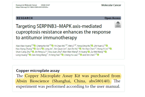
作为深耕生命科学领域的核心供应商,Absin 始终以高品质产品为全球科研工作者提供坚实支撑,赋能各类前沿研究探索。近日,发表于《Molecular Cancer》(文献 DOI:10.1186/s12943-025-02529-x)的一项重磅研究,聚焦胰腺癌铜死亡 resistance 与免疫治疗响应机制,借助 Absin 核心检测工具完成关键实验验证,为胰腺癌治疗提供了全新的靶向策略与理论依据,彰显了 Absin 产品在生命科学研究中的可靠价值。
文献标题: Targeting SERPINB3–MAPK axis-mediated cuproptosis resistance enhances the response to antitumor immunotherapy
发表期刊: Molecular Cancer (IF=33.9)
DOI: 10.1186/s12943-025-02529-x
使用Absin产品: Copper Microplate Assay Kit (abs580140)
聚焦临床痛点,构建多维研究思路
胰腺癌(PDAC)作为恶性程度极高的肿瘤,5 年生存率仅 13%,现有治疗方案疗效有限,亟需挖掘新的治疗靶点与策略。铜死亡作为新型细胞死亡方式,与肿瘤代谢及治疗响应密切相关,但胰腺癌铜死亡 resistance 的调控机制及与免疫治疗的关联尚未明确。
本研究团队围绕这一核心科学问题,构建了 “现象发现 — 机制解析 — 疗法开发” 的完整研究思路:
- 1. 首先通过临床样本分析与细胞实验,验证胰腺癌 “铜累积” 表型与铜死亡 resistance 的关联性;
- 2. 借助多组学分析筛选关键调控因子,明确 SERPINB3 在铜死亡 resistance 中的核心作用;
- 3. 深入解析 SERPINB3 介导铜死亡 resistance 的分子机制,揭示其通过 MAPK 信号通路调控 FDX1 转录的关键路径;
- 4. 针对机制开发 MOF 基纳米递送系统与联合治疗方案,在体内外模型中验证治疗效果。
整个研究逻辑层层递进,从临床现象出发,结合分子生物学、纳米材料学与免疫学等多学科技术,为胰腺癌精准治疗提供了系统性解决方案。
突破性研究成果,重塑胰腺癌治疗认知
本研究通过一系列严谨实验,取得了多项具有里程碑意义的成果:
- 1. 首次发现胰腺癌存在 “铜累积但铜死亡抵抗” 的矛盾表型,且铜含量与肿瘤恶性程度正相关、与患者预后负相关(对应原文图 1B、1F-H);
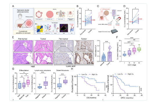
Fig. 1 Pancreatic cancer cells exhibit cuproptosis resistance. A Overall experimental scheme of the study. B Paired line scatter plot showing copper (Cu) concentrations in PDAC tissues (purple dots) (n = 43) and adjacent normal tissues (blue dots) (n = 43). C-D Schematic illustration of cancer cell sorting from cancer tissues of PDAC patients and Cu concentration determination (C). Paired line scatter plot showing the Cu concentrations in the different groups (D). E We used a lipoic acid-specific antibody to quantify the abundance of lipoylated proteins, and the representative images of lipoylated protein IHC in pancreatic cancer tissues (n = 43) and adjacent normal pancreatic tissues (n = 43) from representative patients with PDAC in the cohort from our center are shown (scale bar: 100 μm). Bar chart showing the difference in the average optical density (AOD) values of lipoylated proteins between PDAC tumor and peritumoral tissues. F Box plot showing the Cu concentrations between patients with different AJCC stages. G Statistical graphs of the correlations between Cu concentrations and pathological characteristics in PDAC patients. H Survival analysis of OS and DFS between PDAC patients with high and low Cu concentrations.
- 2. 鉴定 SERPINB3 为胰腺癌铜死亡 resistance 与免疫逃逸的关键调控因子,其高表达与肿瘤进展、不良预后密切相关(对应原文图 1M-N、图 3D-E);
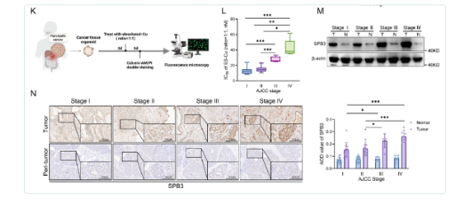
K Schematic illustration of the induction and quantification of cuproptosis in PDOs, which were successfully generated from 32 out of 43 pancreatic cancer specimens obtained from the clinical biobank. L IC50 values for ES-Cu in PDOs with different AJCC stages after 48 h of 2-hour pulsed treatment with different concentrations. M WB analysis showing the expression levels of SERPINB3 in pancreatic cancer tissues and adjacent normal pancreatic tissues from patients with different AJCC stages. N Representative images of SERPINB3 IHC in pancreatic cancer tissues (n = 43) and adjacent normal pancreatic tissues (n = 43) from representative PDAC patients with different AJCC stages in the cohort from our center (scale bar: 100 μm). Box plot showing the AOD values of SERPINB3 between patients with different AJCC stages
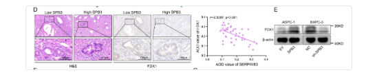
D Representative images of FDX1 immunohistochemical staining in samples with high SERPINB3 expression (SERPINB3hi, n = 22) and low SERPINB3 expression (SERPINB3low, n = 21) pancreatic cancer tissues from representative patients with PDAC in the cohort from our center (scale bar: 100 μm). Scatter plot showing correlation analysis of AOD values between SERPINB3 and FDX1 (right). E WB analysis of FDX1 expression in the indicated ASPC-1 (EV or OE-SERPINB3) and BXPC-3 (NC or sh-SERPINB3) cells.
- 3. 阐明分子机制:SERPINB3 通过抑制 CMA 介导的 MEK1 降解,持续激活 MAPK/ERK 通路,进而抑制 FOXO3 核定位与 FDX1 转录,最终导致铜死亡抵抗;同时上调 PD-L1 表达促进免疫逃逸(对应原文图 4A-E、图 5L-P、图 6A-N);

Fig. 4 SERPINB3 induces cuproptosis resistance by promoting the activation of the MAPK signaling pathway. A The top 7 KEGG terms enriched in genes upregulated after SERPINB3 overexpression in ASPC-1 cells (n = 3 biologically independent sequenced samples per group). B Gene set enrichment analysis of upregulated signatures after SERPINB3 overexpression in ASPC-1 cells (n = 3 biologically independent sequenced samples per group). P values were calculated via Kolmogorov‒Smirnov tests. NES, normalized enrichment score. C The activation status of MAPK signaling pathway-associated proteins was evaluated by a phosphorylation antibody array. D Representative images of ERK1/2 and phosphorylated ERK1/2 immunohistochemical staining in SERPINB3hi (n = 22) and SERPINB3low (n = 21) pancreatic cancer tissues from representative patients with PDAC in the cohort from our center (scale bar: 100 μm). E The ASPC-1-EV and ASPC-1-OE-SERPINB3 cells were treated with or without ES-Cu (40 nM, ratio = 1:1) or trametinib (40 nM) for 24 h, and cell lysates were collected for WB analysis.
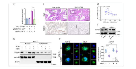
(12) . Correlation analysis of the AOD values for SERPINB3 and FOXO3 (M). N WB analysis of FOXO3 expression in the indicated cells (ASPC-1 [EV or OE-SERPINB3] and BXPC-3 cells [NC or sh-SERPINB3]). O WB analysis of nuclear and cytoplasmic fractions from whole-cell lysates of ASPC-1-EV and ASPC-1-OE-SERPINB3 cells treated with (+) or without (-) trametinib (10 nM) for 24 h. P ASPC-1-EV and ASPC-1-OE-SERPINB3 cells were treated with (+) or without (-) trametinib (100 nM) for 24 h, and representative images of immunofluorescence staining revealed that trametinib reversed the decrease in the expression of FOXO3 in the nucleus induced by the overexpression of SERPINB3 (left). Scatter plot showing the difference in the expression of FOXO3 in the nucleus between the different groups (right)
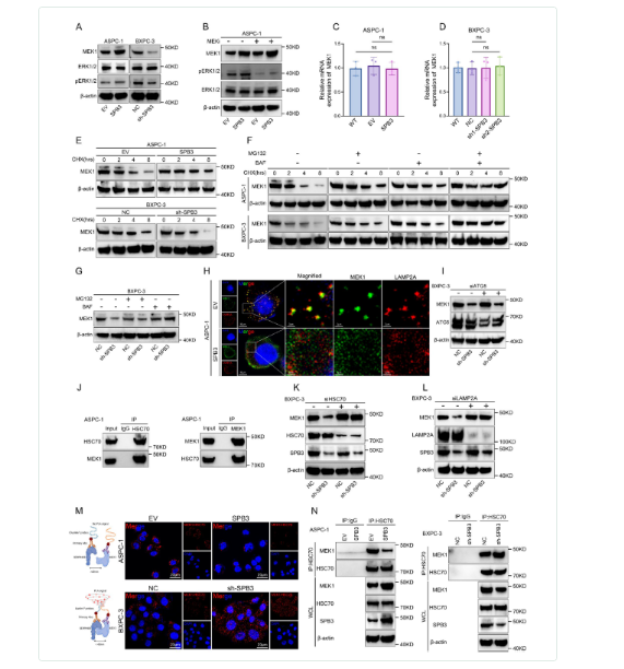
Fig. 6 SERPINB3 inhibits MEK1 degradation through the CMA pathway. A WB analysis of MEK1, ERK1/2 and phosphorylated ERK1/2 expression in the indicated ASPC-1 (EV or OE-SERPINB3) and BXPC-3 cells (NC or sh-SERPINB3). B ASPC-1-EV and ASPC-1-OE-SERPINB3 cells were treated with (+) or without (-) trametinib (100 nM) for 24 h, and cell lysates were collected for WB analysis. C qRT-PCR analysis of MEK1 expression in ASPC-1-EV and ASPC-1-OE-SERPINB3 cells. The data are presented as the means ± SEMs. D qRT-PCR analysis of MEK1 expression in BXPC-3-NC and BXPC-3-sh-SERPINB3 cells. The data are presented as the means ± SEMs. E The indicated ASPC-1 (EV or OE-SERPINB3) and BXPC-3 cells (NC or sh-SERPINB3) were treated with cycloheximide (CHX, 100 μg/ml) for the indicated times, and the resulting cell lysates were collected for WB analysis. F The indicated ASPC-1 (EV or OE-SERPINB3) and BXPC-3 cells (NC or sh-SERPINB3) were treated with CHX (100 μg/ml), MG-132 (10 μM), or BAF (100 nM) for the indicated times, and the resulting cell lysates were collected for WB analysis. G The BXPC-3-NC and BXPC-3-sh-SERPINB3 cells were treated with CHX (100 μg/ml), MG-132 (10 μM), or BAF (100 nM) for 8 h, and cell lysates were collected for WB analysis. H ASPC-1-EV and ASPC-1-OE-SERPINB3 cells were treated with BAF (100 nM) for 3 h, and colocalization between MEK1 (green) and a lysosomal marker (LAMP2A, red) was examined by confocal immunofluorescence staining (scale bar: 10 μm, 2 μm). I BXPC- 3-NC and BXPC-3-sh-SERPINB3 cells were transfected with control siRNA or ATG siRNA for 48 h, and cell lysates were collected for WB analysis. J Wild-type ASPC-1 cells were treated with BAF (100 nM) for 3 h, and cell lysates were collected to detect the interaction between MEK1 and HSC70 by Co-IP and WB analysis. K BXPC-3-NC and BXPC-3-sh-SERPINB3 cells were transfected with control siRNA or HSC70 siRNA for 48 h, and cell lysates were collected for WB analysis. L BXPC-3-NC and BXPC-3-sh-SERPINB3 cells were transfected with control siRNA or LAMP2A siRNA for 48 h, and cell lysates were collected for WB analysis. M Schematic of the PLA for MEK1 and HSC70 (left) and representative fluorescence images (right) of the PLA probe-bound fluorophore in the indicated ASPC-1 cells (top row) and BXPC-3 cells (bottom row) (scale bar: 20 μm). N The indicated ASPC-1 cells (left) and BXPC-3 cells (right) were treated with BAF (100 nM) for 3 h. Cell lysates were collected to detect the interaction between MEK1 and HSC70 via Co-IP and WB analysis. WCL: whole-cell lysate
- 4. 开发 MOF-M-CuSx 纳米递送系统,实现铜离子与 MEK 抑制剂的靶向共递送,联合抗 PD-1 治疗在 SERPINB3 高表达胰腺癌模型中显著抑制肿瘤生长并延长生存期(对应原文图 7J-K、图 9B-E、9M)。
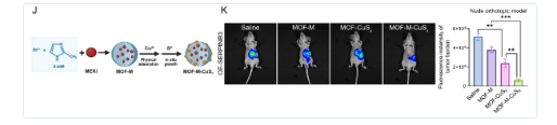
J Schematic illustration of MOF-M-CuSx synthesis. K A nude mouse orthotopic model was generated via the injection of ASPC-1-OE-SERPINB3 cells. Bioluminescence of orthotopic tumors from the mice in different groups treated for three weeks with saline, MOF-M, MOF-CuSx, or MOF-M-CuSx (200 μg/mL, 100 μL, via tail vein) on day 7 after the injection of one million ASPC-1-OE-SERPINB3 cells into the pancreas. Images were collected four weeks after injection. Bar chart showing the fluorescence intensity of the tumor burden in different groups
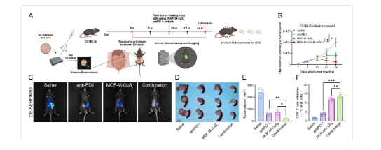
Fig. 9 Induction of cuproptosis in combination with anti-PD-1 therapy can further inhibit the progression of SERPINB3hi PDAC. A The workflow employed to establish C57BL/6 orthotopic tumor models and subsequent treatment. B-C Different groups of C57BL/6 mice received saline, anti-mouse PD-1 monoclonal antibodies (Bio X Cell, 100 μg, i.p., every three days), MOF-M-CuSx (200 μg/mL, 100 μL, via tail vein), or a combination of both for two weeks on day 14 following the orthotopic pancreatic injection of one million KPC-OE-SERPINB3 cells. Each mouse underwent in vivo bioluminescence imaging once a week from day 1 to day 28 postinjection, and the fluorescence intensities representing the tumor burden in each group were plotted individually (B). Representative bioluminescence images of the different groups on day 28 are presented (C). D-E Different groups of C57BL/6 mice received saline, anti-mouse PD-1 monoclonal antibodies (Bio X Cell, 100 μg, i.p., every three days), MOF-M-CuSx (200 μg/mL, 100 μL, via tail vein), or a combination of both for two weeks on day 14 following the orthotopic pancreatic injection of one million KPC-OE-SERPINB3 cells. Tumors were harvested at 4 weeks after pancreatic orthotopic injection
这些成果不仅揭示了胰腺癌铜死亡抵抗的核心机制,更提供了 “铜死亡致敏 + 免疫唤醒” 的新型联合治疗策略,为临床转化奠定了坚实基础。
Absin 核心产品加持,筑牢实验数据基石
在本研究的关键实验环节中,Absin 铜微板检测试剂盒(货号:abs580140) 作为核心检测工具,全程为铜离子浓度定量分析提供支持,成为研究结论可靠输出的重要保障。
该产品采用高特异性检测原理,操作简便、灵敏度高,可精准量化细胞及组织样本中的铜离子浓度,满足临床样本与实验模型的多场景检测需求。其稳定的检测性能与精准的定量结果,完全匹配本研究对铜离子浓度分析的严苛要求,为 “胰腺癌铜累积表型” 的验证提供了直接的实验依据。
关键作用贯穿全程,赋能研究核心环节
Absin 铜微板检测试剂盒(abs580140)在研究中发挥了不可替代的支撑作用,贯穿多个核心实验环节:
- 1. 临床样本验证环节:精准检测 43 对胰腺癌组织与癌旁正常组织的铜离子浓度,证实胰腺癌组织中铜含量显著升高,为 “铜累积表型” 提供关键数据(对应原文图 1B);
- 2. 肿瘤细胞分选验证环节:通过 MACS 分选技术分离胰腺癌组织中的 EpCAM⁺肿瘤细胞与非肿瘤细胞,检测结果明确肿瘤细胞内铜离子浓度显著高于非肿瘤细胞,进一步验证铜累积的细胞特异性(对应原文图 1D);
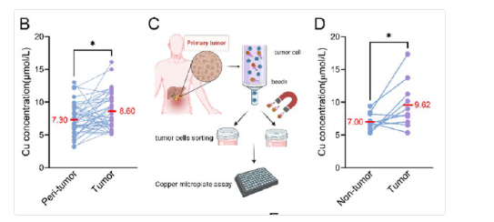
B Paired line scatter plot showing copper (Cu) concentrations in PDAC tissues (purple dots) (n = 43) and adjacent normal tissues (blue dots) (n = 43). C-D Schematic illustration of cancer cell sorting from cancer tissues of PDAC patients and Cu concentration determination
- 3. 治疗效果评估环节:在正交胰腺肿瘤模型中,量化不同治疗方案(ES-Cu、MOF-CuSx 等)对肿瘤组织铜离子浓度的影响,证实 MOF 纳米递送系统可有效提升肿瘤内铜离子富集,为治疗方案的优越性提供直接证据(对应原文图 7I)。
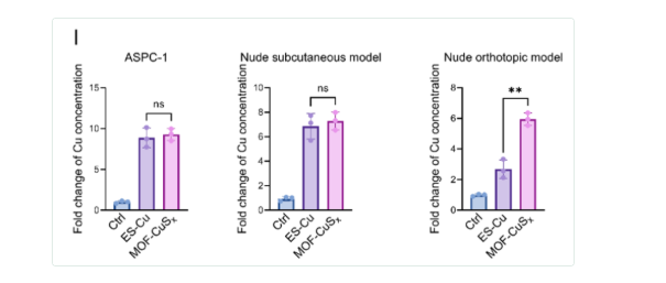
I Bar charts showing the fold change in copper concentration in tumor cells following different drug treatments in pancreatic cancer cells in vitro and in subcutaneous tumors and orthotopic tumors in nude mice.
该产品的高精准性与稳定性,确保了各环节铜离子浓度数据的可靠性,为研究团队揭示铜累积与铜死亡抵抗的关联、验证纳米递送系统的有效性提供了坚实的数据支撑,是研究顺利推进的重要基础。
持续深耕科研需求,助力更多领域突破
本研究成果的发表,再次印证了 Absin 产品在生命科学前沿研究中的核心价值。作为专注于科研需求的供应商,Absin 始终以 “赋能创新研究” 为理念,不断优化产品性能、丰富产品品类,为肿瘤学、分子生物学、免疫学等多个领域提供全方位的实验解决方案。
本文内容基于《Molecular Cancer》(DOI: 10.1186/s12943-025-02529-x)原文献,由 AI 解读整理;文中涉及的原文献图片、数据等知识产权归原期刊及研究团队所有。若存在侵权情形,敬请及时联系我方删除,我方将积极配合处理。