- 移动端


爱必信(上海)生物科技有限公司品牌商
14 年
手机商铺
- NaN
- 0
- 0
- 2
- 2

推荐产品

公司新闻/正文
攻克骨质疏松骨修复难题,微纳羟基磷灰石支架作用机制研究刊发《Bioactive Materials》
17 人阅读发布时间:2026-06-03 11:10
骨质疏松性骨缺损修复一直是临床领域的重大挑战 —— 骨生成能力下降、血管新生紊乱、支架整合效果差等问题,让无数患者面临骨折难愈、残疾风险升高的困境。近期,《Bioactive Materials》发表重磅研究,提出一种 “力学 - 生物活性解耦” 的新型支架设计策略,为骨质疏松骨修复提供了全新解决方案。而 Absin 的优质抗体产品,正是该研究机制验证环节的核心支撑。
文献标题: Hierarchical micro-/nanostructured hydroxyapatite scaffolds promote osteoporotic bone regeneration via activation of hedgehog and HIF-1α signaling
发表期刊: Bioactive Materials (IF=20.3)
DOI: 10.1016/j.bioactmat.2026.01.049
使用Absin产品: Rabbit anti-PTCH1 Polyclonal Antibody (abs115174)
一、创新研究思路:给支架 “定制” 纳米表面,精准激活修复信号
传统羟基磷灰石(HA)支架虽具备骨传导性,但在骨质疏松微环境中,难以同时满足机械支撑与生物活性需求。研究团队另辟蹊径,构建了层级化微纳结构羟基磷灰石(nwHA)支架:以晶须增强羟基磷灰石(wHA)为骨架保障机械强度,再通过真空灌注技术,将五种不同形貌的纳米羟基磷灰石(nHA)涂覆于表面,实现纳米拓扑结构的可编程调控(原文图 1)。
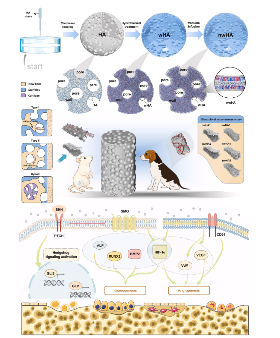
Diagram illustrating the preparation and proposed mechanism of action for nwHA scaffolds. Schematic showing how hierarchical micro-/nanostructured nwHA topographies enhance the osteogenic microenvironment in osteoporotic femoral defects. These topographies promote osteoprogenitor adhesion, vascular infiltration, and morphology-driven ossification modes—including wall-penetrating (type I), surface-appositional (type II), and hybrid patterns—thereby guiding context-specific intramembranous or endochondral bone regeneration.
这种设计的核心巧思在于:将支架的 “力学承载” 与 “细胞调控” 功能分离,通过筛选最优 nHA 形貌,针对性激活骨再生关键信号通路。研究聚焦 Hedgehog 和 HIF-1α 信号通路,系统探究纳米形貌如何影响间充质干细胞(MSCs)成骨分化与血管内皮细胞(HUVECs)血管新生,最终找到骨质疏松微环境的 “精准适配方案”。
二、核心研究成果:nanofiber 涂层支架成最优解,三重突破改写修复格局
经过体外细胞实验与骨质疏松大鼠体内验证,研究取得三大关键发现:
- 1. 形貌决定修复效果:五种 nHA 形貌中,纳米纤维涂层支架(nwHA1)表现最为突出,显著提升骨体积分数、矿化沉积率、机械强度及新生血管密度,其修复效果远超未涂层 wHA 及其他形貌支架(原文图 6、7)。
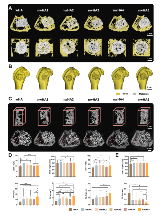
Micro-CT assessment of in vivo bone regeneration at 8 weeks. (A) 3D micro-CT reconstructions showing new bone formation and bone–scaffold interface (white: scaffold; yellow: bone); scale bar: 1 mm. (B) Whole femoral metaphysis reconstruction; scale bar: 2 mm. (C) Top: 3D renderings of trabecular ROI (red dotted box); bottom: corresponding 2D cross-sections at implant–bone interface; scale bar: 1 mm. (D) Quantification of intra-defect bone volume fraction (iBV/iTV), bone mineral density (iBMD), and concentric bone volume (cBV/cTV) within a 100 μm annular ROI; data are mean ± SD (n = 8; ∗p < 0.05, ∗∗p < 0.01, ∗∗∗p < 0.001). (E) Histomorphometric analysis of trabecular microarchitecture parameters including BMD, BV/TV, BS/BV, Conn.Dens, Tb.Th, Tb.Sp, and SMI; data as mean ± SD (n = 8; ∗p < 0.05, ∗∗p < 0.01, ∗∗∗p < 0.001).
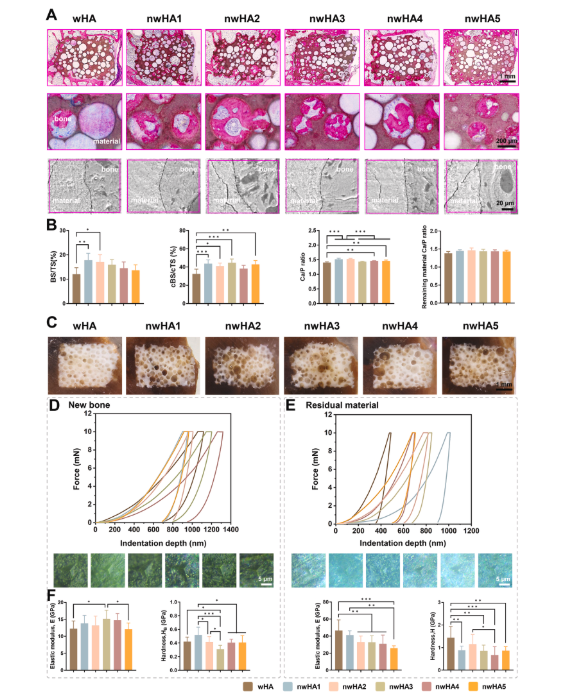
Histological and mechanical characterization of regenerated bone. (A) H&E-stained sections at 4 × and 20 × magnification, and SEM images showing bone–scaffold interfaces; scale bars: 1 mm (4 × ), 200 μm (20 × ), 20 μm (SEM). (B) Quantification of bone surface (BS/TS), contact surface (cBS/cTS) within 100 μm interface zone, and Ca/P ratios (EDS) in newly formed bone vs. residual scaffold; data as mean ± SD (n ≥ 8). (C) Optical imaging of PMMA-embedded undecalcified sections (B: bone, brown; M: material, white); scale bar: 1 mm. (D) Nanoindentation curves, microscopy images, and quantification of elastic modulus and hardness in newly formed bone; scale bar: 5 μm. (E) Corresponding mechanical analysis of undegraded material; all data as mean ± SD (n ≥ 8; ∗p < 0.05, ∗∗p < 0.01, ∗∗∗p < 0.001).
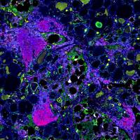
- 2. 解锁三种成骨模式:通过组织学分析,首次明确 nHA 形貌与微环境共同调控三种骨化模式 ——I 型(壁穿透型)、II 型(表面附着型)及混合软骨内 - 膜内成骨模式,其中 nwHA1 更易诱导高效的 I 型骨化(原文图 8)。
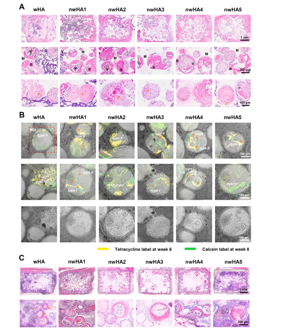
Ossification modes and mineralization dynamics in orthotopic and ectopic models. (A) Decalcified H&E-stained sections at 8 weeks, showing chondrocyte-like clusters at 4 × , 20 × , and 40 × magnification; scale bars: 1 mm (4 × ), 200 μm (20 × ), 100 μm (40 × ). (B) Sequential fluorochrome labeling (tetracycline: red, 6 weeks; calcein: green, 8 weeks) showing type I (wall-penetrating, bridging), type II (surface-appositional, concentric), and hybrid ossification with cartilage-like matrix features. The red dashed rectangle marks the regions of interest (ROI) used for mineral apposition rate (MAR) calculation, which evaluates the kinetics of mineralization by measuring the distance between the two fluorescent labels and dividing by the 14-day interval between the two injections. Scale bar: 100 μm. (C) H&E-stained sections of beagle dorsal muscle implants (12 weeks), demonstrating predominantly type II and hybrid ossification at 4 × and 20 × magnification; scale bars: 1 mm (4 × ), 200 μm (20 × ).
- 3. 信号通路机制明确:nwHA1 通过激活经典 Hedgehog 信号通路(SHH-PTCH1-GLI1 轴),并上调 HIF-1α 表达,在 MSCs 和 HUVECs 中同步促进成骨与血管新生,且两条通路存在双向交叉调控(原文图 3、5)。
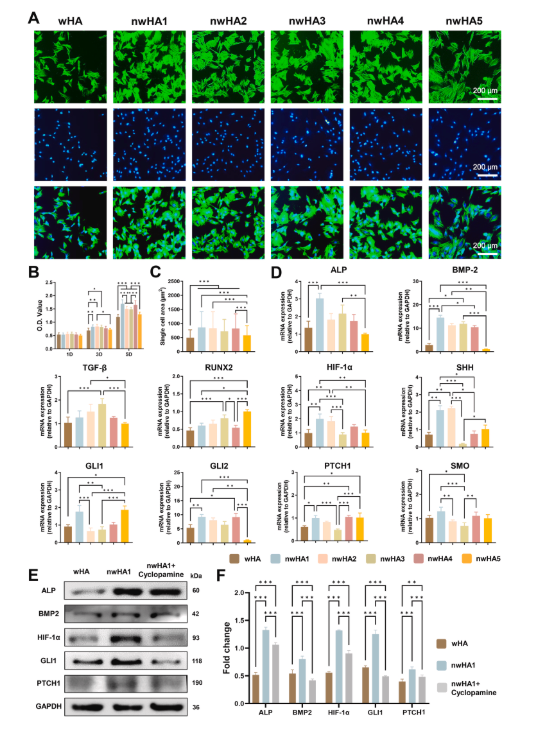
In vitro evaluation of biocompatibility and osteogenic activity of wHA and nwHA scaffolds using MSCs. (A) Fluorescence staining for F-actin (phalloidin-TRITC, green) and nuclei (DAPI, blue), showing enhanced cell spreading. (B) CCK-8 proliferation assay; data presented as mean ± SD (n = 8, ∗p < 0.05, one-way ANOVA with Tukey's post hoc test). (C) Quantification of single-cell area; data presented as mean ± SD (n ≥ 10, ∗p < 0.05, one-way ANOVA with Tukey's post hoc test). (D) qRT-PCR of osteogenic and Hedgehog-related markers (ALP, BMP2, Runx2, HIF-1α, SHH, PTCH1, SMO, GLI1, GLI2) at day 2; data presented as mean ± SD (n = 6, ∗p < 0.05, Kruskal–Wallis with Dunn's post hoc test, when applicable). (E) Western blot and (F) corresponding densitometric quantification of ALP, BMP2, HIF-1α, and GLI1 expression (n > 6, ∗p < 0.05, ∗∗p < 0.01, ∗∗∗p < 0.001, one-way ANOVA with Tukey's post hoc test).
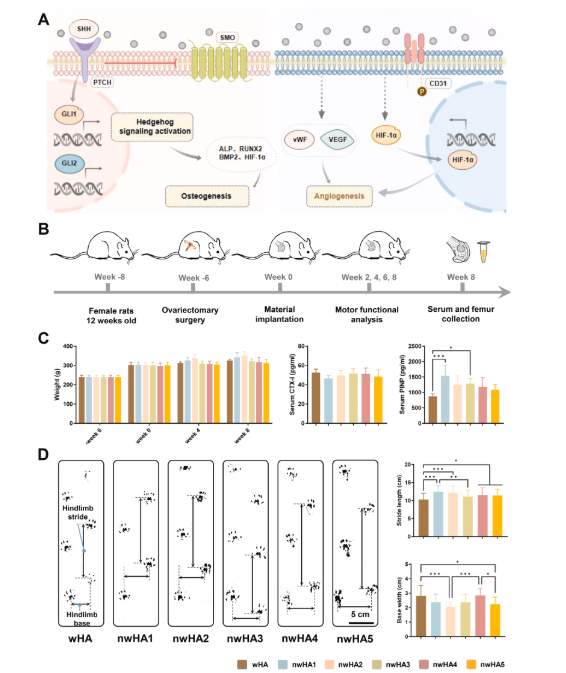
In vivo bone regeneration and functional recovery in osteoporotic rat femoral defects. (A) Schematic illustrating nwHA1-induced osteogenesis via Hedgehog activation in MSCs and pro-angiogenic effects in HUVECs through HIF-1α upregulation. (B) Surgical implantation of wHA and nwHA scaffolds (nwHA1–nwHA5) into distal femoral defects; evaluation at 8 weeks. (C) Body weight and serum markers (CTX-I, PINP) at 8 weeks; data shown as mean ± SD (n = 5; ∗p < 0.05, ∗∗∗p < 0.001, Kruskal–Wallis with Dunn's post hoc test). (D) Gait analysis quantifying stride length and base width as indicators of functional recovery; data shown as mean ± SD (n ≥ 8; ∗p < 0.05, ∗∗p < 0.01, ∗∗∗p < 0.001; one-way ANOVA with Tukey's post hoc test).
三、Absin 产品赋能:PTCH1 抗体精准验证信号通路关键环节
在机制验证的核心实验 ——Western blot 分析中,研究团队选用Absin 的 PTCH1 抗体(货号:abs115174) ,成功完成 Hedgehog 信号通路激活的蛋白水平验证。
Absin 产品在研究中的核心作用:
- 1. 信号通路激活佐证:通过检测 PTCH1 蛋白表达变化,明确 nwHA1 可显著上调 Hedgehog 通路关键分子(包括 PTCH1、GLI1 等),直接证明纳米纤维形貌对通路的激活作用(原文图 3E、3F)。
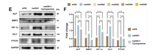
In vitro evaluation of biocompatibility and osteogenic activity of wHA and nwHA scaffolds using MSCs. (E) Western blot and (F) corresponding densitometric quantification of ALP, BMP2, HIF-1α, and GLI1 expression (n > 6, ∗p < 0.05, ∗∗p < 0.01, ∗∗∗p < 0.001, one-way ANOVA with Tukey's post hoc test).
- 2. 机制特异性验证:在环巴胺(Hedgehog 通路抑制剂)干预实验中,Absin PTCH1 抗体清晰显示,抑制剂处理后 PTCH1 蛋白表达显著下调,同步伴随成骨标志物(ALP、BMP2)和 HIF-1α 表达降低,证实 Hedgehog 通路是支架发挥修复作用的核心介导途径。
- 3. 数据可靠性保障:抗体的高特异性与灵敏度,确保了 Western blot 实验中蛋白条带的清晰分辨与定量准确性,为 “通路激活 - 成骨 / 血管新生” 的因果关系提供了坚实数据支撑。
原文图 3E 中,清晰呈现了 wHA 组、nwHA1 组及 nwHA1 + 环巴胺组的 PTCH1 蛋白表达差异,Absin 抗体的精准识别能力让通路调控效应一目了然,成为机制论证的关键视觉证据。
四、行业意义与 Absin 承诺:以优质工具助力骨修复研究升级
该研究的发表,不仅确立了纳米形貌作为骨质疏松骨修复 “可编程调控因子” 的核心地位,更为生物陶瓷支架的临床转化提供了明确方向。而 Absin 作为生命科学研究的可靠合作伙伴,始终以 “提供高品质科研工具” 为使命,其抗体产品凭借高特异性、高灵敏度的优势,已广泛应用于信号通路验证、蛋白表达分析等关键实验场景。
从基础研究到临床转化,Absin 将持续聚焦骨再生、骨质疏松等领域的研究需求,提供包括抗体、试剂、耗材在内的一站式解决方案,助力更多科研团队突破技术瓶颈,推动生命科学研究走向临床应用,为人类健康事业注入强劲动力。
本文内容基于《Bioactive Materials》(DOI: 10.1016/j.bioactmat.2026.01.049)原文献,由 AI 解读整理;文中涉及的原文献图片、数据等知识产权归原期刊及研究团队所有。若存在侵权情形,敬请及时联系我方删除,我方将积极配合处理。